Common Eye Problems in Dogs: 7 Conditions Every Owner Should Recognize
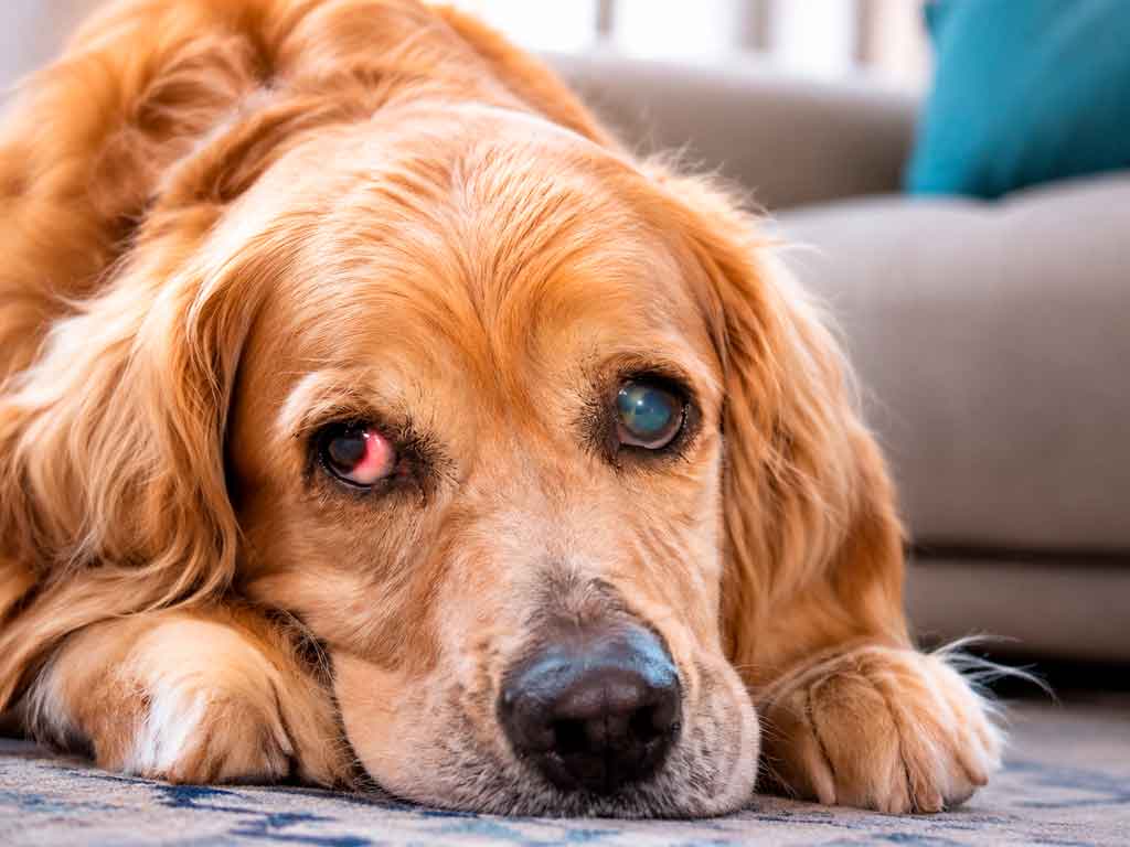
Common eye problems in dogs usually show up as redness, discharge, squinting, or a cloudy surface, and each one carries a different deadline. Cherry eye and mild conjunctivitis can wait a day. Glaucoma and a deep corneal ulcer cannot.
This guide covers the seven conditions veterinarians diagnose most often, the signs that separate a minor irritation from an emergency, and the breeds that carry the highest risk. The information is educational and does not replace an examination, because a dog cannot tell you whether the eye hurts or just itches.
Our articles draw on research and reputable veterinary sources to provide, evidence-based pet care information. This educational content is not a substitute for professional veterinary advice, diagnosis, or treatment. Always consult your veterinarian if your pet develops eye symptoms or its condition worsens.
When common eye problems in dogs need a vet today
Three signs move a dog from “book an appointment” to “go now”: a pupil that stays wide in bright light, a cornea that looks cloudy or blue, and an eye the dog will not open at all. Each one points to pressure, damage, or both.
Dogs hide eye pain well, because the instinct to avoid showing weakness runs deep. A dog that still eats and wags can have a pressure spike or a melting ulcer, so appetite is a poor guide to urgency.
Call your veterinarian immediately, or an emergency clinic after hours, if you notice any of the following.
- The eye looks larger than the other one, or the surface has turned hazy.
- The dog presses its head against furniture or a wall, which often signals pain.
- A yellow or green discharge appears along with a tightly closed eyelid.
- Something has scratched, punctured, or splashed the eye, including shampoo and household cleaners.
- Vision drops suddenly, or the dog bumps into familiar objects.
- The third eyelid slides across the eye and stays there.
The table below compares common visible signs with a reasonable response time. It is a triage aid, not a diagnosis, and a veterinarian may sort the same signs differently after an exam.
| What you see | How fast to act |
|---|---|
| Red eye with clear discharge, dog still eats | Within 24 to 48 hours |
| Pink mass in the inner corner, eye still open | Within a few days |
| Squinting, tearing, or rubbing the face | Same day |
| Cloudy cornea, yellow discharge, eye shut | Emergency, today |
| One eye bulging, pupil wide, head pressing | Emergency, within hours |
| Sudden blindness in a dog that was fine yesterday | Emergency, today |
Some home remedies cause more harm than the original problem. Avoid these while you arrange care.
- Do not use human eye drops, including redness relievers and saline with preservatives.
- Do not apply leftover antibiotics from a previous prescription, because the drug may not match the infection.
- Do not use a steroid drop on a red eye before a vet has stained the cornea.
- Do not remove a collar or cone early, since a single rub can reopen a healing ulcer.
- Do not try to pull a foreign body out with a cotton swab or tweezers.
- Do not wait for a Monday if the eye is closed, cloudy, or bulging.
Clean discharge gently with a warm, damp cloth, working from the inner corner outward, and use a fresh section of cloth for each eye. A cold compress over a closed eyelid can ease comfort for a few minutes, and a cone prevents self-trauma. Neither replaces an exam.
Our guide to dog first aid kit essentials lists the supplies worth keeping at home, including saline for flushing after a chemical splash. If you remember one thing from this article, make it the stain: no drops until the cornea has been checked.
Cherry eye: the pink lump in the corner
Cherry eye happens when the tear gland of the third eyelid slips out of its pocket and swells, according to the Merck Veterinary Manual. Owners describe a round, pink or red mass in the inner corner of the eye.
Young dogs under two years are the typical patients. French bulldogs, English bulldogs, beagles, cocker spaniels, and Boston terriers appear on most breed lists, which is why the French bulldog breed guide is a useful read for new owners.
Owners sometimes mistake the swelling for a tumor or an infection. The tissue itself is normal, it simply sits in the wrong place, and the diagnosis is usually obvious on sight.
The gland produces a large share of the tear film. Removing it solves the cosmetic problem and raises the lifetime risk of dry eye, so most surgeons reposition it instead. Expect a general anesthetic, a recovery collar, and a recheck schedule.
Repair works in most dogs, and a small number need a second procedure. The cone stays on for the full course your vet sets, since one rub against a carpet can undo the sutures.
Cherry eye rarely threatens vision on its own. It does expose the gland to air and dirt, which leads to conjunctivitis and tear film problems when it is left alone for months.
Conjunctivitis: red eye with discharge
Conjunctivitis means inflammation of the pink tissue lining the eyelids. It sits near the top of the list of common eye problems in dogs, and it is also the least specific, because allergies, bacteria, viruses, irritants, and eyelid shape can all produce it.
Clear, watery discharge points toward allergy or irritation, and seasonal pollen is a frequent culprit. Thick yellow or green discharge suggests a bacterial component, often secondary to something else. A dog with a foreign body, a corneal ulcer, or dry eye will look much the same from the outside.
That overlap explains a rule veterinarians repeat: no steroid drop goes into a red eye until a fluorescein stain confirms the cornea is intact. Steroids on an ulcer can accelerate melting and perforation.
Most uncomplicated cases clear within a week of the right topical treatment, along with removal of the underlying cause. A case that rebounds within days usually has something else going on, and the next step is a closer look at tear production, eyelid position, and eyelash abnormalities.
Puppies in group settings pass infectious causes around quickly. A new dog with red, runny eyes should stay home from training class until a vet has looked at them.
Corneal ulcers: the scratch that does not heal
A corneal ulcer is a break in the clear surface of the eye. Trauma, dry eye, entropion, and brachycephalic conformation all contribute, and the last two explain why flat-faced breeds feature so heavily in ophthalmology case loads.
A 2021 study in the Journal of the American Veterinary Medical Association followed 970 brachycephalic dogs referred to ophthalmology services and found corneal ulcers in about 13% of all diagnoses, with dry eye close behind at roughly 10%. Those figures come from a referral population, so they describe the dogs that reached a specialist rather than every dog in the breed.
Owners notice squinting, excessive tearing, redness, and a grey or white haze over the surface. Many dogs paw at the face or rub it along the carpet. A superficial ulcer often heals within a week with topical antibiotics and an Elizabethan collar.
A deep ulcer that thins the cornea can rupture, and that is a surgical emergency. Boxers and some older dogs also develop indolent ulcers, shallow defects that stall because the new tissue will not adhere. Those need debridement or a procedure rather than another week of the same drops.
Chemical burns and plant material cause a share of cases that owners never see happen. A dog that ran through tall grass can carry a seed or a foxtail under the lid, and the scratch keeps reopening until the object is removed.
Glaucoma: pressure that damages the optic nerve
Glaucoma is a rise in pressure inside the eye that damages the retina and optic nerve. It is one of the few eye conditions in dogs that causes genuine, unrelenting pain, and it can take vision within days.
The acute form produces a red eye, a cloudy cornea, a dilated pupil, and an eyeball that feels hard to the touch. Dogs often become withdrawn, stop eating, or press their head into a wall. The Merck Veterinary Manual section on glaucoma describes both the primary inherited form and secondary cases triggered by lens movement, inflammation, or tumors.
Cocker spaniels and basset hounds are classic patients for the inherited form, and several terrier and brachycephalic breeds appear often as well.
Treatment aims to lower pressure fast and protect the second eye, which is usually at risk when the disease is inherited. Drops, oral medication, and surgery all play a role depending on the cause. A dog that reaches a veterinarian within hours has a much better chance of keeping sight than one that arrives after a week of squinting.
Pressure is measured, never guessed. A reading above the normal range supports the diagnosis, and the healthy eye gets checked at the same visit because it often shows early changes first.
Dry eye (KCS): when the eye stops producing tears
Keratoconjunctivitis sicca, usually called dry eye, develops when tear production falls below what the surface needs. Most cases in dogs are immune-mediated, and some medications, nerve damage, and gland removal can contribute. Any dog whose cherry eye gland was removed belongs on a monitoring list.
The classic picture is a dull, lusterless cornea with thick, sticky, mucoid discharge that returns within hours of cleaning. Redness and recurrent infections follow. Unlike a simple irritation, dry eye does not improve on its own and tends to worsen with age.
Cocker spaniels, West Highland white terriers, and shih tzus are overrepresented in practice, which makes breed a useful warning flag during routine checks.
A history of infections that improved with drops and returned within weeks is a common story. Each round of treatment buys comfort without fixing the tear film, and the cornea keeps paying the price.
A Schirmer tear test measures tear production in about a minute. Values below the normal range, combined with clinical signs, support the diagnosis, as the Merck Veterinary Manual entry on KCS explains.
Treatment is lifelong and usually combines a tear stimulant such as cyclosporine or tacrolimus with artificial tears. Owners who keep a simple log of discharge and comfort often notice a flare before the eye looks badly damaged.
Entropion and ectropion: eyelids that point the wrong way
Entropion rolls the eyelid inward, so hair rubs the cornea with every blink. Ectropion rolls it outward, exposing the conjunctiva and letting tears pool in a sagging pocket.
Entropion produces squinting, tearing, and frequent corneal ulcers. It is common in shar peis, chows, Saint Bernards, and English bulldogs, and puppies sometimes need temporary tacking before definitive surgery. Ectropion shows up as a droopy lower lid with chronic redness and discharge, and bloodhounds and cocker spaniels are familiar patients.
Puppies with entropion often receive tacking sutures while the skull grows, and a permanent correction follows once the head shape settles. Waiting on a young dog with recurrent ulcers costs corneal clarity that never comes back.
Signs can appear before the first birthday in predisposed breeds. A puppy that squints outdoors on a sunny day deserves an eyelid check at its next visit, even if the eye looks normal at home.
Both are conformational problems, so drops alone rarely fix them. Surgery corrects the lid, and the earlier it happens in a dog with recurrent ulcers, the less scarring accumulates.
Progressive retinal atrophy: the slow fade
Progressive retinal atrophy, or PRA, is an inherited degeneration of the photoreceptor cells at the back of the eye. It is bilateral, painless, and currently without a cure.
Night blindness usually comes first. Owners notice a dog that hesitates on stairs after dusk, bumps into furniture in dim rooms, or fails to catch a treat tossed in low light. Day vision fades later, and the pupils stay dilated because the retina can no longer respond to light.
The Merck Veterinary Manual section on inherited retinal disease notes that many breeds carry a specific mutation, and DNA tests now identify carriers before breeding. A veterinary ophthalmologist can confirm the diagnosis with a retinal exam.
Breeders can screen stock before a litter is planned, and each DNA test targets the mutation found in that breed. Screening changes the next generation, not the dog in front of you.
No treatment reverses PRA. Management focuses on safety, routine, and mental exercise. Keep furniture in place, use a light on the collar for evening walks, and let the dog lead on familiar routes. Most dogs adapt well when their environment stays predictable.
Which breeds carry the highest risk
Breed matters because conformation, heredity, and grooming needs shape which conditions appear. The table below pairs common breeds with the problems veterinarians watch for first, and it reflects general patterns rather than a prediction for an individual dog.
| Breed | Conditions to watch |
|---|---|
| French bulldog, pug, English bulldog | Corneal ulcers, cherry eye, entropion, dry eye |
| Shih tzu, Lhasa apso | Dry eye, corneal ulcers, cataracts |
| Chihuahua, toy poodle | Cataracts, dry eye, corneal exposure |
| Cocker spaniel | Glaucoma, dry eye, ectropion, cataracts |
| German shepherd | PRA, cataracts, pannus |
| Shar pei, chow chow | Entropion, ectropion, corneal ulcers |
| Golden retriever, Labrador | Cataracts, PRA, eyelid growths |
Most common eye problems in dogs follow breed lines, yet daily care shifts the odds. A shih tzu whose hair falls into the eye will rub the cornea all day, and a weekly trim around the face prevents a surprising number of ulcers.
The pug breed guide covers the routine that reduces exposure for flat-faced dogs, including harness fit, dust control, and eye checks after walks. Our dog health guides collection covers the rest of the body.
Cataracts deserve a mention here because owners often confuse them with PRA. A cataract clouds the lens and can be removed surgically in suitable patients, while PRA affects the retina and cannot. If your dog’s eyes look cloudy rather than dim, the guide to natural cataract treatment for dogs explains what those options actually involve.
How vets tell these conditions apart
A complete exam for common eye problems in dogs takes minutes and answers questions that photos cannot. Expect the veterinarian to work through a short sequence.
- A Schirmer tear test strip measures tear production and screens for dry eye.
- A fluorescein stain highlights corneal scratches and ulcers under blue light.
- Tonometry measures pressure inside the eye and confirms or rules out glaucoma.
- An eyelid and eyelash exam looks for entropion, ectropion, and hairs rubbing the surface.
- A retinal exam with a bright lens assesses the back of the eye for PRA and other disease.
Each test is quick and most dogs tolerate them without sedation. Skipping the stain before dispensing drops is the mistake that turns a small ulcer into a surgical case.
Bring a photo of the eye at its worst, plus a short timeline of when the squinting started. Eyes often look calmer in the clinic than they did at home, and two days of symptoms points somewhere different from two months.
Ask for the stain even when the eye looks better than it did last night. A cornea can appear almost normal while a defect is still open underneath, and the stain settles the question in under a minute.